Imaging technology provides a more clear and comprehensive view of teeth, tissue, nerves and bone inside the mouth.
This is an essential tool for dental practitioners as just an oral examination will not be enough to get a complete observation of a patient’s dental health.
Imaging can uncover conditions that are obstructed from eyesight, such as tooth decay, impacted teeth, bone loss or growths like abscesses, cysts and tumours.
Images can be stored and compared over time to track improvements or changes in potentially adverse areas.
Technological advancements can assist in a more accurate diagnosis and patient understanding, because they can be shown what the dentist is seeing and how important the treatment would be.
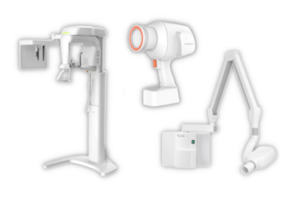
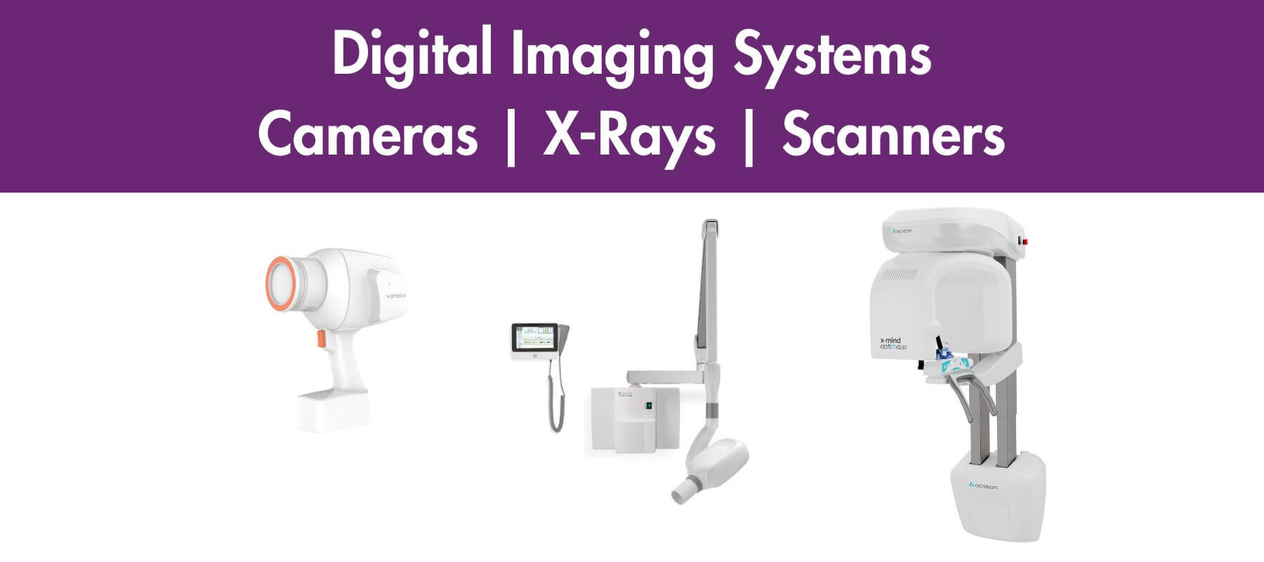
Before digital imaging, dental practices would use film-based radiography that required a dark room to process images. The more modern methods save time, space and resources. The equipment available for dental digital imaging include:
This method is a type of X-ray imaging that uses digital X-ray sensors to replace traditional photographic X-ray film, producing enhanced computer images of teeth, gums and oral structures. Traditional X-rays can also be viewed as digital images by using an X-ray film scanner.
Intraoral X-rays provide excellent detail and are used to detect cavities, check the status of developing teeth and monitor mouth health. Extraoral X-rays are used to detect impacted teeth, monitor jaw growth and development, and identify potential problems between teeth, jaws and facial bones.
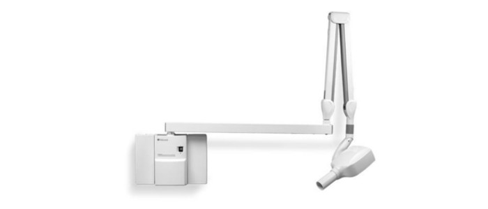
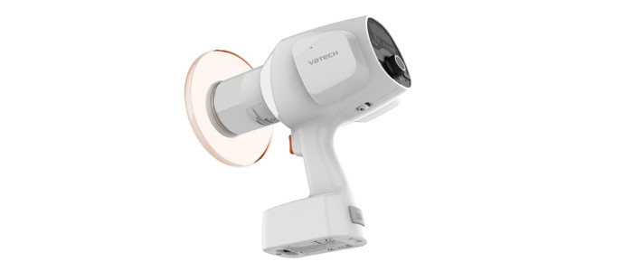
The standard intraoral X-ray device for most dental practices is the wall-mounted, scissor-arm system like the Belmont 097 Belray. Although, the portable and hand-held machines such as the Vatech EzRay Air are gaining popularity and there are advantages to both types.
Bitewing X-rays are used to detect changes in bone density caused by gum disease, decay between teeth, the fit of dental crowns or restorations, and the marginal integrity of tooth fillings.
Periapical X-rays show bone loss around each tooth and are used to detect root structure and surrounding bone structure abnormalities.
Occlusal X-rays reveal the entire arch of teeth and show full tooth development and placement.
When using a conventional wall-mounted unit the operator would leave the room or stand behind a shielded wall during exposure.
Even though the hand-held devices have some very attractive benefits, these fixed units are still popular due to their ease of use, the complete radiation protection and the security of it being attached to a wall, stowed away neatly and permanently connected to power.
If a practice has more than one surgery, there might need to be a wall-mounted unit in each one.
The portability and flexibility of these devices are their main benefit and the patient may feel less intimidated about x-rays due to the operator staying in the room during exposure.
This can also improve efficiency, workflow and ultimately save time. Also, one unit can serve multiple surgeries rather than a fixed unit in each one.
There are many types of extraoral x-rays, their main focus is to detect dental problems in the jaw and skull. They may not provide the detail found with intraoral x-rays, but are integral for identifying issues between the teeth, jaws and temporomandibular joint or other bones around the face.
Types of Extraoral X-Ray:
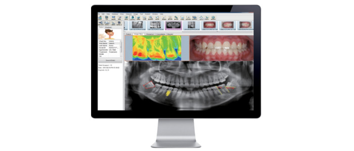
This is a radiographic technique where images are composed of pixels with each value represented by a level on the grey scale, giving a monochromatic picture of the patients’ mouth. 2D or 3D images can be produced, stored or printed and are viewed on screen with several other advantages compared to traditional processes. The benefits of using this system are:
With digital imaging, X-ray machines are still used and require special training for staff. They will also need to be periodically serviced and maintained. Our EclipseCare service plan can ensure that you are CQC compliant, and extend the working life of your equipment.
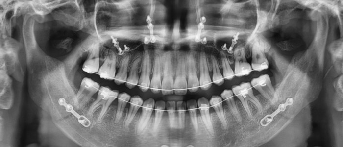
These radiographs produce exceptional images for most dental needs by providing inspection into the internal structure of teeth and supporting bone.
The 2D imaging system we supply is the Vatech PaX-i, which provides the most precise and high-quality panoramic image, improving diagnostic accuracy and increased treatment planning.
In certain cases, 3D imaging might be more appropriate. Computed Tomography (CT) scanners use a narrow fan-shaped beam with multiple exposures around the head to show the internal structures. The 3D image is constructed from a series of 2D images by a cone beam algorithm in the computer software.
This system is called Cone Beam Computed Tomography (CBCT) and can assist in detecting cysts, tumours, infections, maxillofacial injuries and treatment planning for implants and impacted teeth. Other specialist dental applications include orthodontics, cephalometric analysis and periodontics.
Our 3D imaging systems from Vatech provide superior images for panoramic and cephalometric diagnosis:
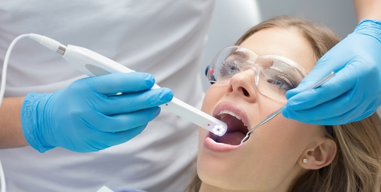
These are essentially small, handheld cameras which produce images not associated with X-rays.
They are a valuable tool for patient education and quick diagnosis. Equipped with an easy capture button to take digital images with wireless or cabled transmission to a screen and a rotatable head to reach anywhere in the oral cavity.
They are ideal to give the patient a tour of their mouth and print images for filing.
Treatments can be more thoroughly discussed and explained, this makes patients more comfortable with their understanding of the procedures. The Intraoral cameras we supply are:
We can inspect any of your X-ray equipment including hand-held devices, usually taking an hour per machine and we recommend an annual service on all of these.
During the service our engineer will check the stability, wear and tear, exposure control, tube-head and fittings. Radiation output and consistency is checked, and documents / paperwork completed.
Call our service team on 01322 421156 or at service@eclipse-dental.com to discuss any service, maintenance or repair needs for your imaging equipment.
The EclipseCare service plans ensure that your practice achieves maximum service uptime; with our operational experience and technical expertise we can help you to run a successful dental practice.
Take a look at this related article – Do I need to have my equipment serviced?
We believe that identifying and delivering the right solution for each practice provides the best value for our customers. Building environments that create better outcomes for dentists and their patients.
Take a look at these case studies to see some of the imaging systems we have installed.
Whether you’re starting from scratch or upgrading your existing surgery, our expert team is here to design, build and equip a practice tailored to your needs.
Fill out this form, and we’ll be in touch within 24 hours to discuss how we can make your project a success.
Eclipse Dental is committed to protecting and respecting your privacy, and we’ll only use your personal information to administer your account and to provide the products and services you requested from us. From time to time, we would like to contact you about our products and services, as well as other content that may be of interest to you.
You can unsubscribe from these communications at any time. For more information on how to unsubscribe, our privacy practices, and how we are committed to protecting and respecting your privacy, please review our Privacy Policy.
I have used them for the first time in recent months for a complicated project in carrying out a practice refurbishment. Eclipse were meticulous and brilliant. Very knowledgable engineers and a beautiful finish to my practice. They really are a family based business who will look after you. Very professional company and will definitely keep using them.
I had my 2 surgery practice completely stripped out and services repositioned with new flooring, new equipment and redecorated. They kept to the agreed time scale and we were up and running in our state-of-the-art new surgery. Thank you Eclipse and I will be using your services again.

I did a lot of research looking for a dental fit-out company before I came across Eclipse Dental. They designed the surgery exactly the way I wanted it to look which suits my requirements perfectly. The final result was phenomenal, just phenomenal!
The engineers that attend our practice are very knowledgeable and always act in a professional manner. If we have an emergency situation John Boyt always tries his best to fit us in. I have no hesitation in recommending Eclipse Dental Engineering to you for all your servicing and breakdown needs.
Eclipse listened to my ideas, they added a lot to them and improved my initial design. They had loads of realistic and creative ideas for a 21st century dental practice! Two surgeries were refurbished on time without any interruption of our clinics.

We would highly recommend the awesome Eclipse team who guided us through the design process, finishing touches and colour schemes. Their ability to combine build works with equipment and dental engineering makes it so much easier and better value.
I am very pleased with the result, it is of a high quality and surpassed my expectations, on the strength of the work done I commissioned some extra wall cabinetry. The whole process was hassle-free and I would be very happy to recommend Eclipse to my friends in the business.
We used Eclipse recently in an emergency as we had equipment failure. They were very quick to respond to our call and were able to get us working again within 2 hours. The engineer was very friendly and professional and I would highly recommend this company.

I would like to express my gratitude to you and your team for your professionalism and prompt response when our dental chair packed up. You attended the same day, removed the damaged chair and installed a rental to allow us to continue practising.
Having dealt with many fit-out companies over the years, Eclipse Dental has been one of the most professional, dependable and sincere companies I have ever worked with. I would not hesitate to use their services again.
Excellent service, Eclipse have worked tirelessly for us and have always come out same day if we have a problem stopping us working. Thanks to all at Eclipse.
Their awareness of CQC regulations regarding equipment relocation and our necessity to minimise downtime was brilliant. We would like to recommend Eclipse Dental to anyone considering a refurbishment or relocation.
Very happy with the service and reliability of the team. From the beginning to the end, everyone was always helpful and very kind. I definitely will recommend Eclipse Dental!
I liked how swiftly the projects were done. Everything was managed, everything was timed and everything was coordinated. And every day we saw something happening. It was very exciting.

The quality of craftsmanship was exceptional—the cabinetry, flooring and all custom elements were made precisely to our specifications and aligned perfectly with our vision for the space.
Brilliant. Just professional! Complete refit of surgery. Flawless.
The boys did well! We didn’t give them much time to prepare but they did a great job, extremely pleased.

We have had the chairs for some time now and are happy with them. They serve what we need them for. The aftercare service is excellent

Eclipse are very flexible and listen to your needs. We are very impressed with the quality of workmanship they delivered. I would not hesitate to continue recommending Eclipse.
Eclipse were always accommodating of any requests and would always go the extra mile. I look forward to working with them for many years to come.

We are delighted with the end result. The practice looks good and, more importantly, works ergonomically and efficiently.